-
 30,000+ Students Sucessfully Trained Annually
-
30,000+ Students Sucessfully Trained Annually

-
30,000+ Students Sucessfully Trained Annually

 30,000+ Students Sucessfully Trained Annually
Advanced Cardiovascular Life Support, commonly known as ACLS, is an advanced emergency response approach for healthcare professionals managing adults with cardiac arrest, acute coronary syndrome, stroke, and other life threatening cardiovascular emergencies. ACLS builds on Basic Life Support and expands clinical skills through cardiac rhythm recognition, advanced airway management, medications, and team-based care.
ACLS uses evidence-based algorithms to guide assessment and treatment according to the patient’s condition. The approach emphasizes early recognition, high-quality CPR, timely defibrillation when indicated, and appropriate care after return of spontaneous circulation
Medication | Clinical Use | Adult Dose and Administration |
Epinephrine | Supports circulation during cardiac arrest in all rhythms. | Administer 1 mg IV or IO every 3 to 5 minutes throughout cardiac arrest. |
Amiodarone | Helps treat VF or pulseless VT that persists after defibrillation. | Deliver 300 mg IV or IO as a bolus; a further 150 mg may be given if necessary. |
Atropine | Helps manage symptomatic bradycardia. | Give 1 mg IV every 3 to 5 minutes, up to a maximum total dose of 3 mg. |
Adenosine | Can terminate certain regular SVTs and restore a normal rhythm. | Administer 6 mg as a rapid IV push; follow with 12 mg after 1 to 2 minutes if required. |
Magnesium Sulfate | Supports treatment of torsades de pointes and hypomagnesemia. | Infuse 1 to 2 g IV, diluted in 10 mL of D5W, over 5 to 20 minutes, according to the clinical indication. |
Dopamine | Provides circulatory support when symptomatic bradycardia or hypotension does not respond adequately to atropine. | Start an IV infusion at 0.05 to 1 mcg/kg/min and adjust according to the patient’s response. |
Norepinephrine | Helps restore blood pressure and perfusion during severe hypotension and shock. | Begin at 0.1 to 0.5 mcg/kg/min, then adjust the infusion rate as clinically indicated. |
Aspirin | Reduces platelet activity in acute coronary syndrome. | Provide 81 to 325 mg orally, preferably chewed when appropriate. |
Nitroglycerin | Relieves ischemic chest discomfort associated with acute coronary syndrome. | Place 0.4 mg sublingually every 5 minutes, with no more than 3 doses. |
Cardiac arrest is managed according to whether the initial rhythm is shockable or nonshockable. Rapid rhythm recognition helps determine whether defibrillation should be delivered or whether CPR and medication therapy should begin immediately.
For ventricular fibrillation or pulseless ventricular tachycardia, defibrillation is the primary treatment. Follow each shock with 2 minutes of CPR and reassess the rhythm, adding medications according to the algorithm.
Asystole and pulseless electrical activity are nonshockable rhythms, so treatment focuses on immediate CPR, epinephrine, and correction of reversible causes.
Bradycardia with a pulse requires evaluation for symptoms, hemodynamic instability, and possible reversible causes. Treatment depends on the patient’s clinical condition and response to initial therapy.
Tachycardia with a pulse is managed based on whether the patient is stable or showing signs of cardiopulmonary compromise. QRS width and rhythm regularity also help determine the appropriate treatment approach.
The Acute Coronary Syndrome algorithm focuses on quickly identifying myocardial ischemia, supporting the patient, and restoring coronary blood flow when needed.
The stroke algorithm focuses on rapid recognition, prompt imaging, and timely treatment to reduce neurological injury.
Rhythm | Description | ECG Appearance |
Normal Sinus Rhythm | Heart rate of 60 to 100 bpm with consistent rhythm intervals. | Evenly spaced QRS complexes with normal morphology. |
Bradycardia | Heart rate of less than 60 bpm in sinus bradycardia. | Widely spaced normal QRS complexes. |
Tachycardia | Heart rate of more than 100 bpm in sinus tachycardia. | Closely spaced normal QRS complexes. |
Atrial Fibrillation (AF) | No distinct P waves with an irregularly irregular ventricular response. | Irregularly irregular rhythm with no clear P waves. |
Ventricular Fibrillation (VF) | Chaotic ventricular electrical activity with no effective cardiac contraction. | Disorganized, irregular waveform. |
Asystole | No detectable cardiac electrical activity. | Flat line after confirming proper lead connection and equipment function. |
Take charge of the resuscitation, provide clear direction, and keep the team focused on the patient's needs.
Share information clearly, give concise instructions, and confirm that important messages are understood.
Give each team member a specific responsibility to maintain an organized response and avoid confusion during care.
Communicate relevant findings, changes in the patient's condition, and treatment updates with the team throughout the resuscitation.
Remain aware of the patient's condition, team performance, treatment progress, and changes that may require action.
Review the response together, identify areas for improvement, and discuss what worked well to strengthen future resuscitation efforts.
A patient collapses, has no pulse, and the monitor shows ventricular fibrillation. What should happen first?
A patient has a slow heart rate accompanied by symptoms or signs of poor perfusion. What is the first-line treatment?
A patient has a rapid heart rate with a pulse. Treatment depends on whether the patient is stable or unstable.
Early recognition and treatment can help reduce neurological injury and improve outcomes in patients with acute stroke.
Identify and treat the Hs and Ts that may be contributing to cardiac arrest or preventing successful resuscitation.
After return of spontaneous circulation, continue close monitoring of airway, breathing, circulation, neurological status, and vital signs to support ongoing stabilization.
Consider patient-specific factors when providing ACLS care, including physiological changes associated with pregnancy and conditions that may affect treatment in older adults.
Verify the medication, dose, route, and timing before administration, while considering contraindications and the patient’s clinical condition.
Use clear communication by giving specific instructions, confirming that the message was understood, and reporting when the task is complete.
Start with the current ACLS algorithms and understand the sequence of assessment and treatment for each emergency.
Know the main ACLS medications, their indications, doses, and timing within each relevant algorithm.
Work through different patient scenarios to strengthen your ability to apply algorithms and make appropriate treatment decisions.
Pay attention to the key points that determine the next intervention, such as rhythm type, patient stability, and response to treatment.
Review algorithms with flowcharts to visualize the sequence of assessment, intervention, and reassessment.
Practice answering questions within a set time to improve your ability to identify important information and make decisions efficiently.
Review important concepts in shorter sessions and revisit them regularly to strengthen recall and understanding.
A few key details can help you understand what to expect from an ACLS course and certification.
Strong BLS skills are required before progressing through ACLS training, including high-quality CPR, ventilation, and defibrillation.
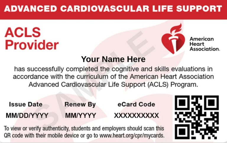
Complete all required course components and meet the applicable assessment requirements to receive ACLS certification.
ACLS certification is generally valid for 2 years before renewal is required.
Course time can vary based on the training format and provider, with completion taking approximately 8.25 to 18.5 hours.
Knowledge, skills, and a calm approach can help healthcare providers respond effectively during emergencies.

EDUCATIONAL USE ONLY:
This content is intended for educational use only. It does not replace an official ACLS course, current ACLS algorithms, local protocols, or professional clinical judgment.
Yes. A strong understanding of Basic Life Support is expected before enrolling in an ACLS course. BLS skills such as high-quality CPR, AED use, and basic airway management form the foundation for advanced cardiovascular care.
ACLS certification is generally valid for two years. Renewing your certification helps maintain current knowledge of resuscitation guidelines and reinforces the skills needed to manage cardiovascular emergencies.
Many training providers offer blended learning that combines online coursework with an in-person skills evaluation. Course availability and training formats may vary by certification provider.
Basic Life Support focuses on recognizing life threatening emergencies, performing high-quality CPR, using an AED, and providing basic airway support. Advanced Cardiovascular Life Support builds on these skills by introducing advanced airway management, ECG rhythm interpretation, emergency medications, and evidence-based treatment algorithms.
If your ACLS certification expires, you may be required to complete a renewal or provider course, depending on your employer’s policies and the certification provider’s requirements. Renewing your certification helps keep your knowledge and clinical skills up to date.
No. ACLS is required only for healthcare professionals whose roles involve managing cardiovascular emergencies or providing advanced resuscitation care. Certification requirements vary depending on the employer and clinical setting.