-
 30,000+ Students Sucessfully Trained Annually
-
30,000+ Students Sucessfully Trained Annually


-
30,000+ Students Sucessfully Trained Annually

 30,000+ Students Sucessfully Trained Annually
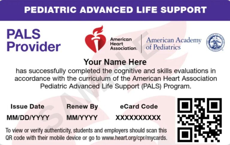
Prepare for your American Heart Association PALS certification with a complete review of pediatric emergency care. Learn how to recognize respiratory failure, shock, and cardiac arrest while mastering assessment tools, treatment algorithms, rhythm recognition, medications, and effective team communication.
Pediatric Advanced Life Support is an advanced resuscitation program that teaches healthcare professionals how to recognize early signs of deterioration and manage critically ill or injured pediatric patients. The course combines systematic assessment, high-quality CPR, airway management, rhythm recognition, pharmacology, and team-based resuscitation.
Unlike adult emergencies, pediatric patients often show subtle signs before their condition becomes critical. PALS helps providers recognize these warning signs early and respond using a structured approach that supports accurate clinical decisions and consistent care throughout the resuscitation process.
A severe choking emergency requires different techniques for infants and children.
Repeat 5 back blows followed by 5 heel-of-hand chest thrusts. Do not perform abdominal thrusts. If the infant becomes unresponsive, begin CPR without checking for a pulse. Remove only a visible object from the mouth and never perform a blind finger sweep.
Repeat 5 back blows followed by 5 abdominal thrusts. If the child becomes unresponsive, begin CPR without checking for a pulse. Remove only a visible object from the mouth and never perform a blind finger sweep.
Strong team dynamics improve coordination, reduce delays, and help deliver high-quality resuscitation. Every team member should communicate clearly, understand their responsibilities, and work together toward the same goal.
Each team member should have a defined responsibility, such as managing the airway, performing chest compressions, administering medications, or documenting events. Clear role assignments improve efficiency and reduce confusion.
Recognize your level of training and experience. Ask for assistance when a task exceeds your scope of practice and allow qualified team members to perform advanced interventions.
Speak up when you identify a potential error or patient safety concern. Respectful communication helps prevent mistakes and supports better patient care.
Share important patient information, assessment findings, and treatment updates with the entire team. Open communication keeps everyone informed and improves decision-making.
Regularly summarize the patient's condition, interventions completed, and the team's progress. Frequent reassessment helps identify changes and guides the next steps in care.
Repeat verbal instructions to confirm they were heard correctly, and acknowledge when tasks have been completed. This communication method reduces misunderstandings during resuscitation.
Treat every team member with professionalism and respect. A positive team environment encourages collaboration, effective communication, and high-quality patient care.
The Pediatric Assessment Triangle is a rapid visual assessment tool used to identify critically ill or injured infants and children. A complete assessment takes only a few seconds and focuses on three key areas that help determine the child’s overall condition.
Appearance reflects the child’s neurological status and overall well-being. Evaluate the following:
Assess the effort required for the child to breathe and look for signs of respiratory distress.
Evaluate skin color and perfusion to identify signs of poor circulation.
The ABCDE approach provides a structured method for identifying and treating life-threatening conditions in infants and children. Assess each component in order and address any critical problems before moving to the next step.
Assess the effectiveness of circulation.
Perform a quick neurological assessment.
Perform a complete physical assessment while protecting the child from heat loss.
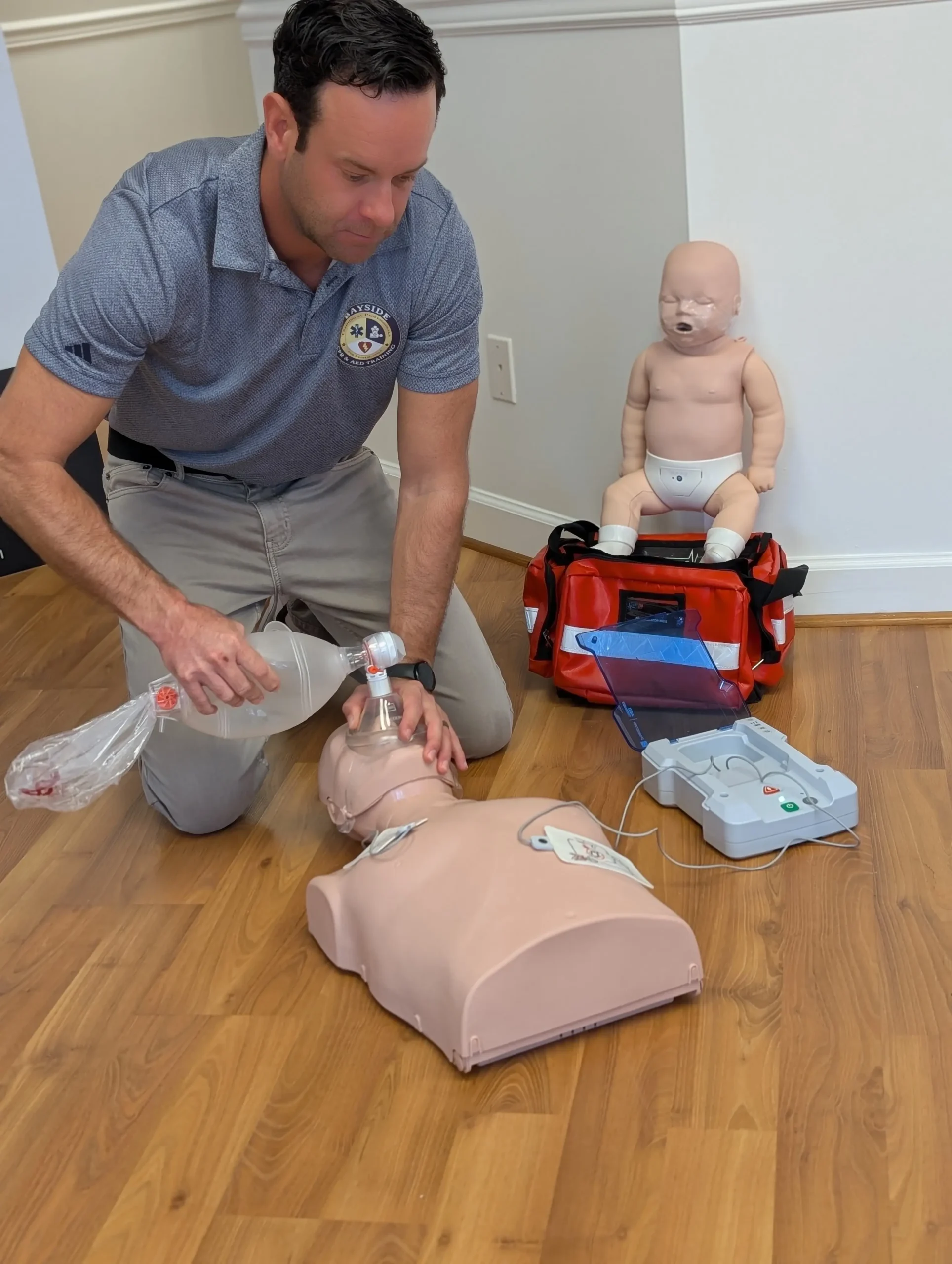
Targeted actions help improve outcomes after return of spontaneous circulation (ROSC) and support safe, coordinated pediatric care. Ongoing monitoring, careful medication use, and effective communication remain essential throughout post-resuscitation management.
After return of spontaneous circulation (ROSC), monitor and manage the patient’s temperature to prevent fever. Maintain a temperature of 37.5°C or lower to reduce the risk of additional neurological injury.
Provide enough oxygen to maintain an oxygen saturation between 94% and 99% while avoiding both hypoxemia and hyperoxia. Adjust oxygen delivery based on the patient’s condition and monitoring results.
Continue care after ROSC with ongoing monitoring and support of breathing, circulation, and neurological function. Identify and treat the underlying cause of the cardiac arrest while supporting recovery.
Verify the correct medication, dose, concentration, and route of administration before giving medications. Accurate medication management helps prevent errors during critical care.
Communicate instructions clearly during resuscitation and confirm understanding by having the receiver repeat the message. This closed-loop communication helps ensure that tasks are performed correctly.
Pediatric resuscitation requires adjustments based on the patient’s age and size. Airway equipment, compression techniques, ventilation, and medication doses should always be selected according to the child’s developmental stage and body size.
An infant is a child younger than 1 year of age, excluding newborns in the delivery setting. Use infant-specific airway equipment, perform chest compressions using the appropriate infant technique, and calculate medication doses based on the infant’s weight.
A child is 1 year of age until the onset of puberty. Select equipment based on the child’s size, adjust compression technique as appropriate, and use weight-based medication dosing to provide safe and effective treatment.
Once immediate life-threatening conditions have been managed, perform a secondary assessment to gather additional information and identify injuries or medical conditions that may require treatment.
The SAMPLE history provides important details about the child’s condition and medical background.
Perform a targeted physical examination based on the child’s condition and suspected illness or injury.
Early recognition of respiratory problems allows timely treatment and may prevent respiratory arrest. Distinguishing respiratory distress from respiratory failure helps guide appropriate interventions.
Respiratory distress occurs when a child is working harder to breathe but is still able to maintain oxygenation and ventilation.
Respiratory failure occurs when a child cannot maintain adequate oxygenation and ventilation.
Act quickly when signs of respiratory compromise appear. Open and maintain the airway, remove secretions with suction if needed, provide oxygen, and assist breathing when the child is unable to maintain effective ventilation.
Different respiratory conditions affect different parts of the respiratory system. Identifying the likely cause helps determine the most appropriate treatment priorities.
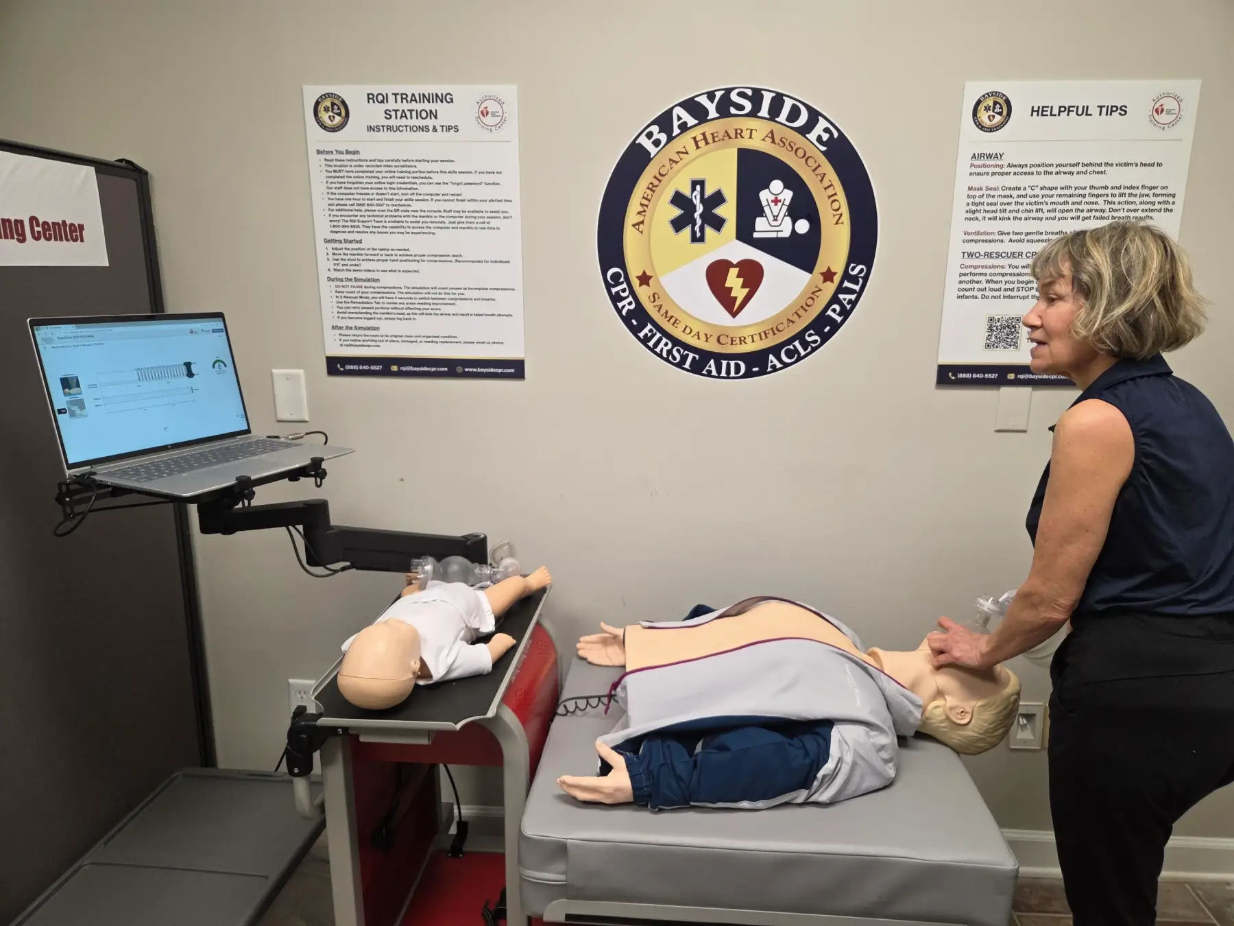
Shock in children can progress quickly, so early recognition and intervention are important. Evaluate circulation, provide support based on the child’s condition, and reassess after each intervention.
Look for signs of poor perfusion such as abnormal heart rate, weak pulses, delayed capillary refill, cool or mottled skin, and changes in mental status. Remember that low blood pressure is a late sign of pediatric shock.
Provide oxygen and ventilation support when needed, establish IV or IO access, administer fluids or medications as indicated, and treat the underlying cause of shock.
Monitor the child’s response by evaluating heart rate, perfusion, mental status, blood pressure, and oxygenation. Adjust interventions based on the child’s condition and continue frequent reassessment.
Recognizing the type of shock helps guide treatment and supports timely interventions. Although the causes differ, all forms of shock require prompt assessment, continuous monitoring, and frequent reassessment.
After each fluid bolus, reassess the child’s response and monitor for signs of fluid overload.
Care does not end after return of spontaneous circulation (ROSC). Ongoing management focuses on protecting vital organs, preventing secondary injury, and supporting recovery through continuous monitoring and timely interventions.
Monitor core temperature in comatose patients and prevent fever by maintaining a temperature of 37.5°C or lower. Maintain systolic and mean blood pressure above the 10th percentile for the child’s age and sex to support adequate perfusion.
After cardiac arrest, oxygen and ventilation should be adjusted to keep SpO₂ between 94% and 99%, avoid both hypoxemia and hyperoxemia, and maintain carbon dioxide levels within the normal range or the child’s baseline.
Treat seizures promptly, evaluate neurological outcomes using a multimodal and time-based approach, and provide rehabilitation planning with continued follow-up after recovery.
Description: Normal sinus rhythm is an organized rhythm with a heart rate appropriate for the child’s age. Sinus bradycardia occurs when the heart rate is lower than the expected range for a child’s age and is commonly caused by low oxygen levels (hypoxia).
Management: Assess airway, breathing, and circulation and correct the underlying cause. Provide oxygen and ventilation support as needed. If heart rate remains below 60/min with poor perfusion despite effective oxygenation and ventilation, start CPR and follow the Pediatric Bradycardia Algorithm. Use epinephrine for persistent compromise and atropine only for increased vagal tone or primary AV block.
Description: Sinus tachycardia occurs when the heart rate is faster than the expected range for the child’s age. The rhythm usually develops gradually and has normal P waves. Common causes include fever, pain, low blood volume, hypoxia, and anxiety.
Management: Assess airway, breathing, and circulation. Treat the underlying cause, provide oxygen or other support when needed, and monitor the child’s response to treatment.
Description: Supraventricular tachycardia (SVT) is a rapid heart rhythm that usually starts suddenly and has a fixed rhythm pattern. P waves may be absent or appear abnormal. SVT is commonly suspected when the heart rate is 220/min or higher in infants and 180/min or higher in children.
Management: Assess airway, breathing, and circulation. For stable patients, try vagal maneuvers first, followed by adenosine if needed. For patients with poor perfusion or signs of instability, perform synchronized cardioversion starting at 0.5 to 1 J/kg. Increase the dose to 2 J/kg if the initial dose is not effective. Adenosine may also be given when IV or IO access is available.
Description: Ventricular tachycardia is a rapid ventricular rhythm that requires immediate assessment for the presence of a pulse and signs of instability.
Management: Check for a pulse first. If the child has a pulse but is unstable or has signs of poor perfusion, use synchronized cardioversion. Begin with 0.5 to 1 J/kg. If the rhythm continues, increase the energy dose to 2 J/kg. If no pulse is present, treat as a shockable cardiac arrest rhythm.
Description: Ventricular fibrillation and pulseless ventricular tachycardia are shockable cardiac arrest rhythms that require rapid treatment.
Management: Deliver defibrillation as soon as possible and resume CPR immediately after each shock. Give epinephrine after the second shock, or earlier if defibrillation cannot be performed quickly. Consider amiodarone or lidocaine when VF or pulseless VT continues despite treatment.
Description: Asystole and pulseless electrical activity (PEA) are nonshockable cardiac arrest rhythms that do not respond to defibrillation.
Management: Continue high-quality CPR, give epinephrine as soon as possible, and repeat every 3 to 5 minutes. Identify and treat reversible causes. Do not attempt defibrillation for asystole or PEA.
Management depends on whether the cardiac arrest rhythm is shockable or nonshockable. Identify the rhythm promptly and follow the appropriate PALS algorithm while providing high-quality CPR and minimizing interruptions.
Check breathing and pulse together within 10 seconds or less. Start CPR if there is no pulse or if the heart rate remains below 60/min with signs of poor perfusion despite effective oxygenation and ventilation.
Without an advanced airway, provide 30 compressions to 2 breaths with one rescuer and 15 compressions to 2 breaths with two rescuers. Give each breath over about 1 second and provide only enough air to produce visible chest rise.
2025 Pediatric Basic Life Support Guidelines.
2025 Pediatric Advanced Life Support Guidelines.
2025 Pediatric Resuscitation Algorithms.
2025 Course Updates and Pediatric Advanced Life Support Provider Manual.