-
 30,000+ Students Successfully Trained Annually
-
30,000+ Students Successfully Trained Annually


-
30,000+ Students Sucessfully Trained Annually

 30,000+ Students Successfully Trained Annually
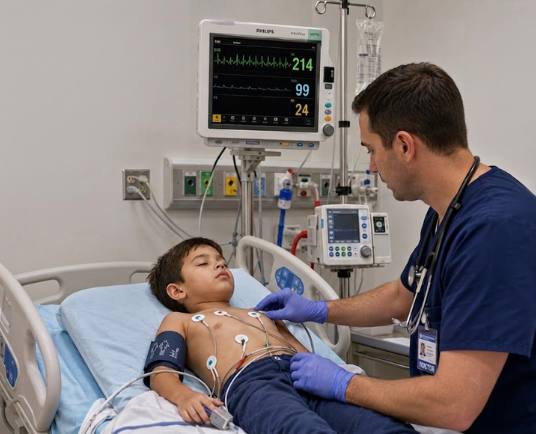
When a child’s heart starts beating much faster than normal, every moment is crucial. Knowing how to recognize the warning signs and respond quickly can make a significant difference in the child’s outcome. Pediatric tachyarrhythmia with a pulse is a rapid heart rhythm that occurs while the heart is still producing a detectable pulse. This condition can reduce the heart’s ability to pump blood effectively, especially when the heart rate is extremely high or remains elevated for a prolonged period. Common signs include a rapid heartbeat, palpitations, chest discomfort, dizziness, difficulty breathing, poor feeding in infants, altered mental status, and low blood pressure. Some children remain stable, while others develop signs of poor perfusion that require immediate medical attention. Prompt recognition is essential because untreated tachyarrhythmias can lead to serious complications.
The Pediatric Tachyarrhythmia With a Pulse Algorithm, developed by the American Heart Association and the American Academy of Pediatrics as part of Pediatric Advanced Life Support (PALS), provides a step-by-step approach to evaluating and treating children with a rapid heart rhythm. It helps healthcare providers determine whether the child is stable or unstable, identify the type of tachyarrhythmia, and select the most appropriate treatment. Depending on the child’s condition, the algorithm may recommend supportive care, vagal maneuvers, medications such as adenosine, or synchronized cardioversion for unstable patients. By following this structured approach, healthcare teams can make timely decisions, improve coordination during emergencies, and increase the chances of safely restoring a normal heart rhythm.
Note: This pediatric tachyarrhythmia with a pulse algorithm guideline remains valid for the period 2025–2030.
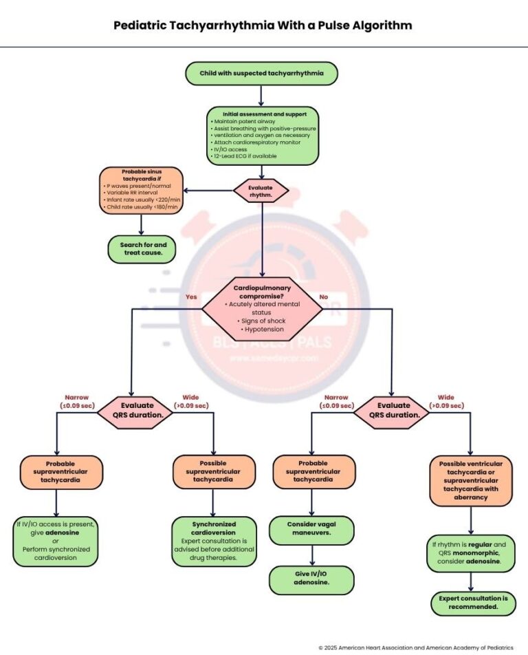
The first step is to identify a child who may have tachyarrhythmia, which is an abnormally fast heart rhythm. A child may have a very rapid pulse along with symptoms such as palpitations, chest discomfort, dizziness, trouble breathing, poor feeding in infants, or changes in alertness. Early recognition is important because some fast heart rhythms are harmless, while others require immediate medical treatment to prevent serious complications.
Begin by making sure the child's airway is open and clear. If the child is not breathing well, assist breathing with positive pressure ventilation and provide oxygen as needed. Attach a cardiorespiratory monitor to continuously check the heart rate and rhythm. Establish IV or IO access so medications can be given quickly if necessary. If available, obtain a 12 Lead ECG to help identify the type of tachyarrhythmia and guide the next steps in treatment.
Carefully evaluate the child's heart rhythm to determine the most likely cause of the rapid heart rate. If the rhythm suggests probable sinus tachycardia, continue to Step 4 for further assessment. If the rhythm shows signs of cardiopulmonary compromise, move directly to Step 6 because the child may need urgent treatment to restore a stable heart rhythm.
Sinus tachycardia is likely when normal P waves are present, the RR interval varies, the infant's heart rate is usually less than 220 beats per minute, and the child's heart rate is usually less than 180 beats per minute. These findings suggest that the fast heart rate is a normal response to another medical condition rather than an abnormal heart rhythm. After confirming these features, continue to Step 5.
When sinus tachycardia is suspected, focus on finding and treating the underlying cause instead of treating the heart rate alone. Common causes include fever, pain, dehydration, anxiety, low oxygen levels, infection, or blood loss. Correcting the underlying problem usually allows the heart rate to return to normal without additional rhythm-specific treatment.
Assess the child for signs of cardiopulmonary compromise, including an acutely altered mental status, signs of shock, or hypotension. These findings indicate that the fast heart rhythm is affecting blood circulation and requires prompt treatment. If any of these signs are present, continue to Step 7. If the child remains stable without these findings, proceed to Step 12.
Measure the QRS duration on the ECG to determine whether the rhythm is narrow or wide. If the QRS is narrow, measuring less than or equal to 0.09 seconds, continue to Step 8. If the QRS is wide, measuring greater than 0.09 seconds, proceed to Step 10 because wide complex tachycardias often require a different treatment approach.
Step 8: Probable Supraventricular Tachycardia
A narrow QRS complex in a child with cardiopulmonary compromise often suggests supraventricular tachycardia. This rapid rhythm usually begins above the ventricles and can reduce the heart's ability to pump blood effectively. Once this rhythm is recognized, continue to Step 9 for immediate treatment.
If IV or IO access is available, administer adenosine because it can stop many types of supraventricular tachycardia and restore a normal rhythm. If adenosine cannot be used or the child needs immediate treatment, perform synchronized cardioversion. This procedure delivers a controlled electrical shock that is synchronized with the heartbeat to safely restore a normal rhythm.
A wide QRS complex with cardiopulmonary compromise may indicate ventricular tachycardia. This rhythm starts in the ventricles and can quickly become life-threatening if not treated promptly. Once ventricular tachycardia is suspected, continue immediately to Step 11.
Perform synchronized cardioversion to restore a normal heart rhythm in a child with unstable ventricular tachycardia and a pulse. Before giving additional antiarrhythmic medications, seek expert consultation whenever possible. Specialist guidance helps ensure the safest and most appropriate treatment for the child.
If the child does not have cardiopulmonary compromise, evaluate the QRS duration to guide treatment. A narrow QRS measuring less than or equal to 0.09 seconds leads to Step 13. A wide QRS measuring greater than 0.09 seconds leads to Step 16 because different types of tachyarrhythmias require different management.
In a stable child, a narrow QRS complex commonly indicates supraventricular tachycardia. Although the child is stable, treatment is still needed to prevent symptoms from worsening. Continue to Step 14 to begin simple measures that may restore a normal rhythm.
Begin by considering vagal maneuvers, which stimulate the vagus nerve and may slow the heart rate or stop supraventricular tachycardia. These techniques are simple, noninvasive, and are often tried before medications when the child remains stable. After attempting vagal maneuvers, proceed to Step 15.
If vagal maneuvers are unsuccessful or not appropriate, administer adenosine through an IV or IO line. Adenosine briefly blocks electrical conduction through the heart, allowing many episodes of supraventricular tachycardia to return to a normal rhythm. Continue to monitor the child's response after the medication is given.
A wide QRS complex in a stable child may represent ventricular tachycardia or supraventricular tachycardia with aberrant conduction. Because these conditions can appear similar on an ECG, careful evaluation is important before selecting treatment. Continue to Step 17 for the next decision.
If the rhythm is regular and the QRS complex is monomorphic, consider giving adenosine. In selected cases, adenosine may help identify the rhythm or successfully treat supraventricular tachycardia with aberrancy. After this assessment and treatment, continue to Step 18.
After the initial evaluation and treatment, obtain expert consultation to guide further care. A pediatric cardiologist or another qualified specialist can recommend additional testing, medications, or advanced treatment if needed. Ongoing monitoring and expert advice help ensure the child receives the safest and most effective care.
This section explains the key points in the sidebar using simple and easy-to-understand language. It highlights the main signs of probable supraventricular tachycardia and reviews the recommended treatment doses to help you understand the pediatric tachyarrhythmia algorithm more clearly.
Supraventricular tachycardia has several signs that help healthcare providers recognize this fast heart rhythm. Understanding these features makes it easier to identify the condition and choose the right treatment.
1. P waves absent or abnormal
Missing or abnormal P waves may indicate that the rapid heart rhythm is not starting from the heart’s normal pacemaker. This finding is common in supraventricular tachycardia.
2. RR interval not variable
A fixed RR interval means the heart beats at a very regular rate. This steady pattern is often seen in supraventricular tachycardia.
3. Infant rate usually greater than or equal to 220 per minute
A heart rate of 220 beats per minute or higher in an infant strongly suggests supraventricular tachycardia. This is faster than the heart rate usually seen with sinus tachycardia.
4. Child rate usually greater than or equal to 180 per minute
A heart rate of 180 beats per minute or higher in a child may indicate supraventricular tachycardia. The child’s symptoms and ECG findings should also be evaluated.
5. History of abrupt rate change
A sudden start or stop of a fast heart rate is a common feature of supraventricular tachycardia. This pattern helps distinguish it from sinus tachycardia, which usually speeds up and slows down gradually.
Correct dosing helps treat pediatric tachyarrhythmia safely and effectively. Follow the recommended dose and method for each treatment to improve the chance of restoring a normal heart rhythm while reducing the risk of complications.
1. Synchronized Cardioversion
Begin synchronized cardioversion with 0.5 to 1 Joule per kilogram. If the first shock does not work, increase the energy to 2 Joules per kilogram, and provide sedation if needed without delaying treatment.
2. Adenosine IV or IO Dose
Give adenosine at 0.1 milligrams per kilogram as a rapid IV or IO push, followed immediately by an IV flush, with a maximum first dose of 6 milligrams. If needed, give a second rapid dose of 0.2 milligrams per kilogram followed by an IV flush, without exceeding the maximum dose of 12 milligrams.
In short, a well-organized approach is essential when treating pediatric tachyarrhythmia with a pulse because quick decisions can improve outcomes and reduce complications. By following each step of the algorithm, healthcare providers can identify the type of fast heart rhythm, recognize signs of instability, treat the underlying cause when appropriate, and deliver the right intervention at the right time. Understanding the differences between sinus tachycardia, supraventricular tachycardia, and ventricular tachycardia helps guide safe and effective care. Regular practice with the algorithm also builds confidence, strengthens teamwork, and prepares healthcare professionals to respond calmly during emergencies. With prompt assessment and timely treatment, many children can achieve a stable heart rhythm and receive the care they need as quickly as possible.
Take the next step in your emergency care training with PALS certification at Same Day CPR. Gain practical skills that help you provide safe and effective care for children in critical situations.