-
 30,000+ Students Successfully Trained Annually
-
30,000+ Students Successfully Trained Annually


-
30,000+ Students Sucessfully Trained Annually

 30,000+ Students Successfully Trained Annually
No parent, caregiver, or healthcare provider ever expects to witness a child suddenly collapse. Yet when it happens, fear and uncertainty can overwhelm everyone present. In those critical moments, knowing how to recognize cardiac arrest and respond immediately can make the difference between life and death. Pediatric cardiac arrest is a life-threatening emergency that occurs when a child’s heart suddenly stops pumping blood effectively, causing blood flow to the brain and other vital organs to stop. Unlike adults, cardiac arrest in infants and children is often caused by breathing problems, severe infections, drowning, trauma, or other conditions that lead to a lack of oxygen rather than a primary heart problem. Without immediate treatment, cardiac arrest can quickly result in permanent brain damage or death, making rapid recognition and intervention essential.
The Pediatric Cardiac Arrest Algorithm is a structured, evidence-based approach developed by the American Heart Association (AHA) and the American Academy of Pediatrics to guide healthcare providers in the resuscitation and management of cardiac arrest in infants and children. It outlines the sequence of critical actions, including recognizing cardiac arrest, starting high-quality CPR, assessing heart rhythm, delivering defibrillation when appropriate, administering medications, and identifying reversible causes. By following the algorithm, medical teams can respond quickly and consistently, reduce errors during high-pressure situations, and improve the chances of restoring circulation and achieving better survival and neurological outcomes.
Note: This pediatric cardiac arrest algorithm guideline remains valid for the period 2025–2030.

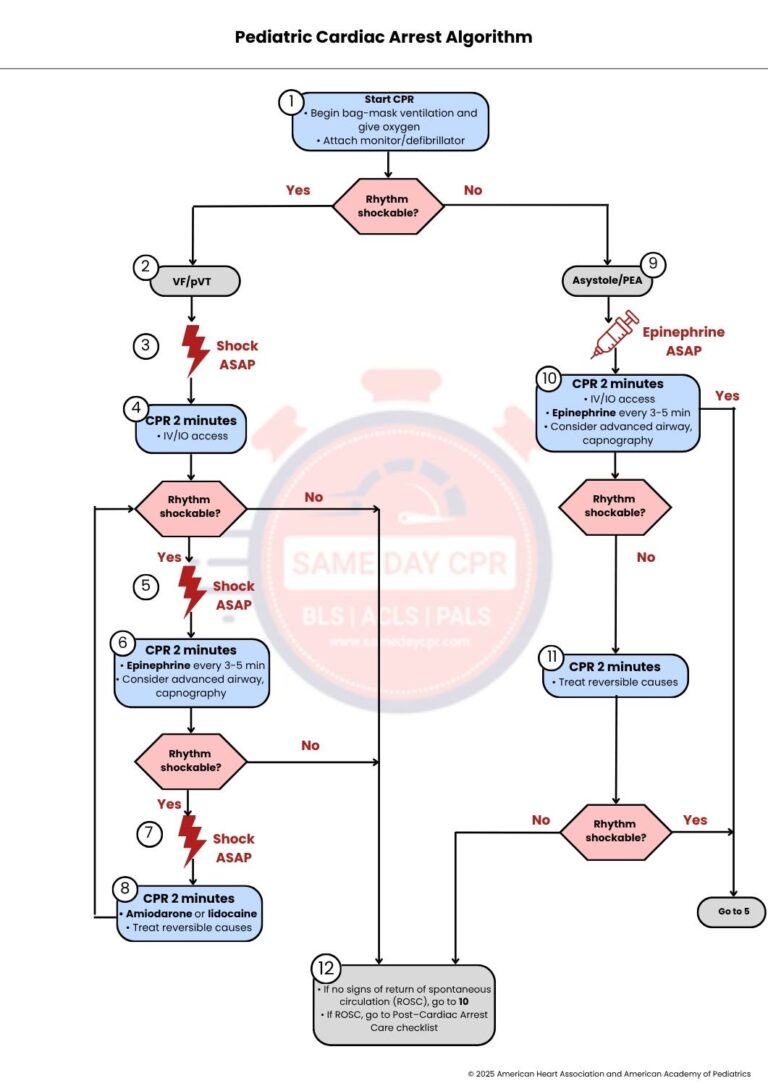
When a child has a cardiac arrest, quick action can make a big difference. This step-by-step guide explains what to do at each stage so you can respond with confidence and give the best possible care.
Start CPR (Cardiopulmonary Resuscitation) immediately when cardiac arrest is recognized. Begin high-quality chest compressions while another rescuer provides bag-mask ventilation with oxygen to help deliver oxygen to the lungs and body. As soon as possible, attach a monitor or defibrillator to identify the patient's heart rhythm. After checking the rhythm, determine whether it is shockable. If the rhythm is shockable, proceed to Step 2. If the rhythm is nonshockable, proceed to Step 9.
If the monitor shows ventricular fibrillation (VF) or pulseless ventricular tachycardia (pVT), the patient has a shockable cardiac rhythm. These rhythms require immediate defibrillation because the heart is beating in a disorganized way and cannot pump blood effectively. Once VF or pVT is identified, proceed directly to Step 3.
Deliver a shock with the defibrillator as soon as possible (ASAP). Early defibrillation is one of the most effective treatments for shockable cardiac arrest because it can stop the abnormal heart rhythm and allow the heart to return to a normal rhythm. After the shock is delivered, immediately continue with Step 4 without delaying CPR.
Perform CPR continuously for 2 minutes after the shock to maintain blood flow to the brain and other vital organs. During this time, establish intravenous (IV) or intraosseous (IO) access so that medications can be administered if needed. After 2 minutes of CPR, reassess the heart rhythm. If the rhythm is shockable, proceed to Step 5. If the rhythm is nonshockable, proceed to Step 12.
Deliver another shock as soon as possible if the rhythm remains shockable. Repeated defibrillation may be necessary to restore a normal heart rhythm. After delivering the shock, continue immediately with Step 6 to minimize interruptions in chest compressions.
Continue CPR for 2 minutes while administering epinephrine every 3 to 5 minutes to improve blood flow during resuscitation. If appropriate, consider placing an advanced airway and use capnography to monitor ventilation and assess the effectiveness of CPR. After 2 minutes, reassess the rhythm. If it is shockable, proceed to Step 7. If it is nonshockable, proceed to Step 12.
If the heart rhythm is still shockable, deliver another shock as soon as possible. Rapid defibrillation remains the recommended treatment for persistent VF or pVT. After the shock is given, immediately continue with Step 8.
Continue CPR for 2 minutes after the shock. During this cycle, administer an antiarrhythmic medication such as amiodarone or lidocaine to help manage persistent shockable rhythms. At the same time, identify and treat any reversible causes of cardiac arrest, such as low oxygen levels, electrolyte imbalances, or other correctable conditions. After 2 minutes, reassess the rhythm. If it remains shockable, return to Step 5. If it is nonshockable, proceed to Step 12.
If the patient's rhythm is asystole or pulseless electrical activity (PEA), these are nonshockable rhythms, so defibrillation is not indicated. Instead, administer epinephrine as soon as possible (ASAP) while continuing high-quality CPR. After giving epinephrine, continue with Step 10.
Continue CPR for 2 minutes while establishing IV or IO access if it has not already been obtained. Administer epinephrine every 3 to 5 minutes, and consider placing an advanced airway with capnography to monitor ventilation. After completing 2 minutes of CPR, reassess the heart rhythm. If it becomes shockable, proceed to Step 5. If it remains nonshockable, proceed to Step 11.
Continue CPR for another 2 minutes while identifying and treating any reversible causes that may be preventing the heart from restarting. Correcting these underlying problems can improve the chances of successful resuscitation. After 2 minutes, reassess the rhythm. If it becomes shockable, proceed to Step 5. If it remains nonshockable, proceed to Step 12.
At this stage, assess whether the patient has achieved return of spontaneous circulation (ROSC), meaning the heart has started pumping blood effectively again. If there are no signs of ROSC, return to Step 10 and continue the nonshockable cardiac arrest algorithm. If ROSC is achieved, move to the Post–Cardiac Arrest Care checklist to provide ongoing treatment, stabilize the patient, and improve recovery.
This section explains the key principles of pediatric cardiac arrest management. It summarizes high-quality CPR techniques, defibrillation shock energy, medication doses, advanced airway management, and the reversible causes of cardiac arrest to provide a quick reference during learning or emergency care.
High-quality CPR gives the best chance of keeping blood moving until the heart starts beating again. Using the right technique can help protect the brain and other organs while emergency care continues.
Shock energy is an important part of treating a heart rhythm that can respond to a defibrillator. Using the correct amount of energy can help restore a normal heartbeat while reducing the risk of harm.
Medicines can support CPR by helping the heart respond better to treatment. Giving the correct dose at the right time is an important part of pediatric cardiac arrest care.
1. Epinephrine IV or IO dose: 0.01 mg/kg
Epinephrine helps improve blood flow to the heart and brain during cardiac arrest. Give 0.01 mg/kg using the 0.1 mg/mL concentration by the IV or IO route, with a maximum single dose of 1 mg.
2. Amiodarone IV or IO dose: 5 mg/kg bolus
Amiodarone is used for shock-resistant ventricular fibrillation or pulseless ventricular tachycardia. Give 5 mg/kg as a bolus, up to a maximum of 300 mg per dose, and it may be repeated for up to 3 doses if needed.
3. Lidocaine IV or IO dose: 1 mg/kg
Lidocaine is another medicine that can treat certain life-threatening abnormal heart rhythms. It may be used instead of amiodarone at a dose of 1 mg/kg IV or IO.
Advanced airway care helps keep air moving into the lungs during CPR. Using the right device and checking its placement can improve oxygen delivery and support effective resuscitation.
1. Endotracheal intubation or supraglottic airway
An advanced airway helps keep the airway open and allows oxygen to reach the lungs during resuscitation. This can be done by placing an endotracheal tube or a supraglottic airway device.
2. Use ETCO₂ to confirm and monitor endotracheal tube placement
End-tidal carbon dioxide, called ETCO₂, confirms that the endotracheal tube is in the trachea and not the esophagus. It also helps monitor the quality of ventilation and the effectiveness of CPR.
Some health problems can lead to cardiac arrest in children, but many of them can be found and treated quickly. Learning these reversible causes helps caregivers and health workers act fast and improve the child’s chance of recovery.
| H’s (Reversible Physiologic Causes) | T’s (Reversible Mechanical/Toxin Causes) |
|---|---|
| Hypovolemia: A severe loss of blood or body fluids can reduce blood flow and stop the heart from pumping well. | Tension pneumothorax: Air trapped around the lung can squeeze the heart and make it hard to pump blood. |
| Hypoxia: Low oxygen levels can prevent the heart and brain from working properly. | Tamponade, cardiac: Fluid around the heart can press on it and stop it from filling and pumping normally. |
| Hydrogen ion (acidosis): Too much acid in the body can weaken the heart and affect its normal rhythm. | Toxins: Harmful substances can damage the heart or interfere with its normal function. |
| Hypoglycemia: Very low blood sugar can reduce the energy the body needs to keep the heart working. | Thrombosis, pulmonary: A blood clot in the lungs can block blood flow and strain the heart. |
| Hypokalemia or hyperkalemia: Potassium levels that are too low or too high can cause dangerous changes in the heartbeat. | Thrombosis, coronary: A blood clot in the heart arteries can cut off blood supply and stop the heart from working properly. |
| Hypothermia: A very low body temperature can slow the heart and reduce its ability to function. |
In short, the pediatric cardiac arrest algorithm provides a clear and organized approach for responding to one of the most serious emergencies in children. Every step, from starting CPR to giving medicines and treating the cause, plays an important role in improving the child’s chance of survival and recovery. Acting quickly, following the correct sequence, and working well as a team can make a life-saving difference during these critical moments. Regular training and practice also help healthcare professionals stay prepared and respond with confidence. By understanding and applying this algorithm, medical teams can deliver safe, timely, and effective care when every emergency occurs.
At Same Day CPR, we provide PALS courses that give healthcare professionals the knowledge and hands-on skills needed to recognize pediatric cardiac arrest, respond with confidence, and deliver high-quality care in real emergencies.