-
 30,000+ Students Successfully Trained Annually
-
30,000+ Students Successfully Trained Annually

-
30,000+ Students Sucessfully Trained Annually

 30,000+ Students Successfully Trained Annually
When someone goes into cardiac arrest, the resuscitation team has only a few minutes to respond effectively. The ACLS algorithm was created for exactly this situation. It turns decades of evidence from resuscitation science into a clear step-by-step process that trained providers can follow under intense pressure. The goal is to make sure life-saving actions, like defibrillation, airway support, giving medications, and identifying the underlying cause, are done in the correct order and without delay.
This guide explains what the ACLS algorithm is, how it is organized, and the main pathways every certified provider should understand. It also covers the key medications used and highlights important updates from the American Heart Association (AHA) 2025 CPR & ECC Guidelines.
It’s meant to serve as a practical reference and orientation resource, not a substitute for official AHA certification training or your hospital’s clinical protocols.
Advanced Cardiovascular Life Support (ACLS) refers to a set of clinical treatments and decision-making pathways used to manage cardiac arrest, life-threatening arrhythmias, acute coronary syndrome, and stroke. The “algorithm” itself is the structured, flowchart-style guidance published by the AHA. It lays out an organized sequence of “if this, then that” steps to help providers decide what to assess, what actions to take, and when to escalate care.
Rather than being a single pathway, ACLS is actually a collection of related algorithms. Each one is designed for a specific emergency, but they all share the same core principles: delivering high-quality CPR, performing early defibrillation when appropriate, carrying out a systematic patient assessment, and identifying reversible causes.
In practice, the provider’s role is to quickly determine which algorithm fits the patient’s condition and then carry it out efficiently as part of a coordinated resuscitation team.
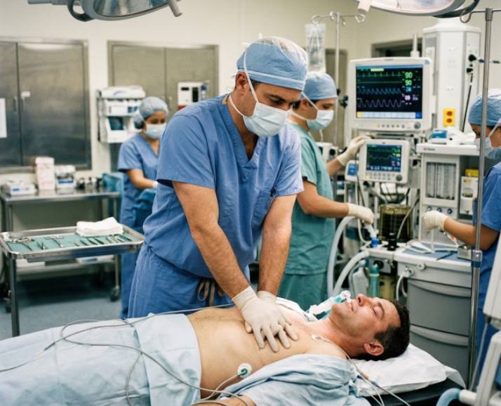
Every ACLS algorithm is built on the same core structure. Providers follow this sequence to stay organized and ensure no critical step is missed during a high-pressure emergency.
The process begins with the Basic Life Support (BLS) assessment: check responsiveness, activate the emergency response system, and assess breathing and pulse at the same time (this should take no longer than 10 seconds). If no pulse is detected, high-quality CPR is started immediately. A defibrillator or AED is applied as soon as it becomes available.
This stage introduces more advanced interventions. The airway is secured if necessary, and correct placement is confirmed using quantitative waveform capnography when an advanced airway is in place. IV or IO access is established, cardiac monitoring leads are attached, and vital signs are obtained. The focus here is on stabilizing the patient while gathering essential real-time data.
The Primary Assessment of ACLS: ABCDE: Airway–Breathing–Circulation–Disability–Exposure
The team then moves to a more detailed evaluation, often guided by the SAMPLE history: Signs and symptoms, Allergies, Medications, Past medical history, Last meal, and Events leading up to the arrest. This is combined with a focused physical exam and a structured search for reversible causes, commonly remembered as the “H’s and T’s.”
Once these foundational steps are completed, the patient’s heart rhythm or clinical presentation determines which specific ACLS algorithm is followed next.
Every ACLS algorithm depends on one constant in the background: high-quality CPR. Without it, no medication or advanced intervention can be effective enough on its own. This means maintaining consistent, effective compressions with as few interruptions as possible:
In short, everything in ACLS is built on the foundation of doing CPR well and doing it consistently.
This algorithm guides resuscitation for pulseless patients, integrating high-quality CPR, rhythm checks every two minutes, and timely defibrillation for shockable rhythms. It directs epinephrine and antiarrhythmic administration, emphasizes minimizing interruptions in chest compressions, and considers reversible causes (H's and T's) throughout the resuscitation effort.
This algorithm addresses symptomatic bradycardia with a pulse, typically a heart rate below 50 bpm, causing hypotension, altered mental status, or chest pain. It prioritizes identifying underlying causes, administering atropine as first-line treatment, and escalating to transcutaneous pacing, dopamine, or epinephrine infusions when atropine proves ineffective.
This algorithm manages rapid heart rhythms with a pulse, distinguishing stable from unstable presentations. Unstable patients with hemodynamic compromise require immediate synchronized cardioversion. Stable patients are further classified by QRS width and regularity, guiding decisions between vagal maneuvers, adenosine, or antiarrhythmic medications.
This algorithm guides management immediately following return of spontaneous circulation, focusing on optimizing oxygenation, blood pressure, and ventilation. It emphasizes targeted temperature management, 12-lead ECG acquisition, treating precipitating causes, and coordinated critical care to improve neurological outcomes and survival.
This algorithm guides rapid recognition and treatment of suspected heart attacks, emphasizing early ECG acquisition, aspirin administration, and risk stratification. It directs decisions regarding reperfusion therapy, including timely transfer for percutaneous coronary intervention or fibrinolytics, based on ECG findings and symptom onset time.
This algorithm adapts standard resuscitation for pregnant patients in cardiac arrest, emphasizing left lateral uterine displacement to relieve aortocaval compression, standard hand placement for compressions, and continuous fetal monitoring considerations. It also addresses preparation for emergency cesarean delivery if return of spontaneous circulation isn't achieved promptly.
This algorithm streamlines rapid stroke identification and treatment, emphasizing time-sensitive assessment using tools like the Cincinnati Prehospital Stroke Scale. It guides immediate CT imaging, blood glucose checks, and eligibility evaluation for fibrinolytic therapy or thrombectomy, since "time is brain" throughout the care pathway.
“Note: AHA hasn’t updated the Acute Coronary Syndrome and ACLS Suspected Stroke Algorithm in 2025, so they still follow the 2020 guidelines.”
These are the key medications commonly used within ACLS algorithms. It’s important to always double-check exact dosages with your most recent AHA course materials and follow your institution’s protocols, as recommendations can be updated between guideline cycles.
| Drug | Primary Use | Typical Adult Dosing |
|---|---|---|
| Epinephrine | Cardiac arrest (all rhythms), symptomatic bradycardia (infusion) | 1 mg IV/IO every 3–5 minutes during arrest |
| Amiodarone | Refractory VF/pulseless VT | 300 mg IV/IO first dose, 150 mg IV/IO second dose |
| Lidocaine | Alternative to amiodarone for refractory VF/pulseless VT | 1–1.5 mg/kg IV/IO first dose |
| Atropine | First-line for symptomatic bradycardia | 1 mg IV every 3–5 minutes, up to a defined maximum |
| Adenosine | Stable, regular, narrow-complex tachycardia (SVT) | 6 mg rapid IV push, then 12 mg if needed |
| Magnesium sulfate | Torsades de pointes | Per weight-based protocol |
| Dopamine | Second-line for symptomatic bradycardia (infusion) | Titrated infusion |
A defibrillator and medications can only help if they’re treating the actual cause of the arrest. That’s why ACLS providers use the “H’s and T’s” as a quick checklist during PEA or asystole, and in any cardiac arrest that isn’t responding to standard treatment. It helps the team step back and ask: What is actually causing this?
| H’s – Reversible Physiologic Causes | T’s – Reversible Mechanical/Toxin Causes |
|---|---|
| Hypoxia: Insufficient oxygen delivery to tissues, resulting in impaired cardiac performance. | Tension pneumothorax: Air becomes trapped in the pleural space, causing lung collapse and compressing the heart and major vessels. |
| Hypovolemia: Significant loss of blood or fluids leading to reduced circulating volume and decreased cardiac output. | Cardiac tamponade: Fluid buildup in the pericardial sac that restricts the heart’s ability to pump effectively. |
| Hydrogen ion (acidosis): Elevated acidity in the bloodstream that disrupts normal cellular and heart function. | Toxins: Poisoning or drug effects that impair normal cardiac or respiratory activity. |
| Hyperkalemia / Hypokalemia: Abnormal potassium levels that can trigger serious cardiac rhythm disturbances. | Coronary thrombosis: A clot obstructing the coronary arteries, leading to myocardial infarction (heart attack). |
| Hypothermia: Unusually low body temperature that slows down metabolic processes and heart function. | Pulmonary thrombosis (pulmonary embolism): A clot blocking pulmonary arteries, reducing blood flow to the lungs and compromising oxygen exchange. |
The ACLS algorithm shows what needs to be done, but good team dynamics determine whether it actually gets done effectively in an emergency.
Successful resuscitation depends on clear communication and well-defined roles. Each team member knows their job—whether it’s doing chest compressions, managing the airway, establishing IV/IO access, giving medications, leading the code, or documenting the event.
Teams also rely on closed-loop communication, where instructions are repeated back to confirm they were heard and understood. Members are expected to speak up if something doesn’t look right, and to stay within their role while supporting others as needed.
This is a key reason the AHA places so much emphasis on team training. In practice, knowing the algorithm is only part of the picture—how well the team works together often has just as much impact on patient outcomes.
The ACLS certification is needed for healthcare workers who handle serious heart emergencies in clinical settings. It prepares them to respond quickly and work together during critical situations.
ACLS certification is typically valid for two years. Training is usually offered in two formats: a blended option that combines online coursework with an in-person skills session, or a fully in-person course. In both cases, it’s important to make sure the program follows the most current AHA guidelines before you enroll.
If your certification card is still valid, you don’t need to retake the course immediately just because new guidelines have been released. However, when it’s time for renewal, it’s a good idea to choose a course that reflects the latest updates. Most training providers update their online modules and ACLS megacode scenarios to match the newest algorithm changes and recommendations.
In summary, the ACLS algorithm gives healthcare teams a clear path to follow when time matters most. By combining strong CPR, quick defibrillation, the right medications, and a steady search for the root cause, providers can act with confidence instead of guessing under pressure. Learning these steps builds skill, but lasting success comes from practicing them with a group that communicates well and trusts one another. Whether you are new to resuscitation or simply renewing your certification, staying familiar with this approach helps you stay calm in a crisis and gives your patients the best possible chance at survival.
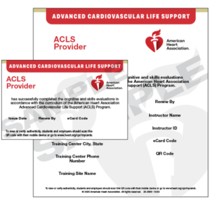
Want to enroll in ACLS certification? Same Day CPR provides quick and convenient lifesaving training prepared to fit your schedule. Begin with a short online course, then complete a short in-person skills session at one of our CPR Verification Stations. Once both components are finished, you’ll receive your American Heart Association ACLS Provider card the same day and be better prepared and confident to act in critical situations.